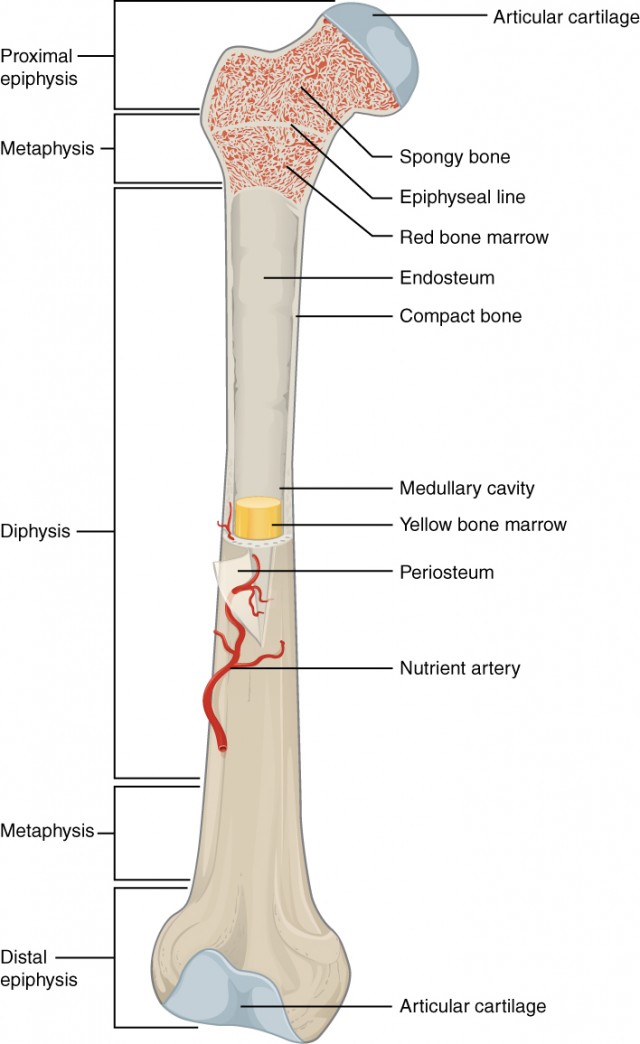
Bone Cross Section Diagram - Tissue: A&P 141 LAB - Anatomy & Physiology 1014 with Idk ... : Diagram of a cross section of the coiled cochlea.. Diagram with articular cartilage, marrow, medullary cavity and periosteum. Diagram with articular cartilage, marrow, spongy bone, medullary cavity, endosteum, diaphysis, and periosteum. They are similar to the topographic profiles that you created in the topographic maps chapter, but they also show the rock types and geologic structures. A cross section of a human long bone. (micrograph provided by the regents of university of michigan.
Explaned distal and proximal epiphysis. Bone cross section diagram ipad folio cases. In a cross section of a bone we can see two types of bone tissue: Schematic drawing of a longitudinal section through a. Healthy tooth diagram isolated on white background vector.
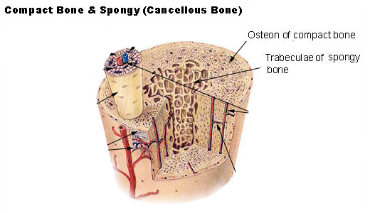
In a cross section of a bone, you can usually see two ... from useruploads.socratic.org 850 x 1270 png 173kb. Compact bone is the outer layer and the spongy bone forms the inner layer. Please will you consider sharing with me? (b) in this micrograph of the osteon, you can clearly see the concentric lamellae and central canals. Explaned distal and proximal epiphysis. Diagram with articular cartilage, marrow, spongy bone, medullary cavity, endosteum, diaphysis, and periosteum. Crosssection cutaway diagram dry cell battery. (micrograph provided by the regents of university of michigan.
From wikimedia commons, the free media repository.
Diagram of a cross section of the coiled cochlea. The centroidal locations of common cross sections are well documented, so it is typically not necessary to calculate the location with the equations above. It consists of two layers; They build the entire picture, improve your understanding, consolidate the information and facilitate recall. Cross section of bone diagram.
Bone Structure | Anatomy and Physiology I from s3-us-west-2.amazonaws.com For example, to read this diagram literally, since the cartilage can be seen inside the cutaway section of bone, it. Diagram with articular cartilage, marrow, medullary cavity and periosteum. Cross section of the human retina. 850 x 1270 png 173kb. Hope you enjoy and please. Explaned distal and proximal epiphysis. Vector illustration scheme of bone cross section. Explaned distal and proximal epiphysis.
Cross section diagram of human bone, bone, cross section diagram of human bone.
Each system contains haversian canals surrounded by concentric lamellae of bone tissue 48. This is a short tutorial using blender 2.8 that shows how to create a bone cross section and using images to create the textures. Vector illustration scheme of bone cross section. Fermur bone with labels and diagram. (micrograph provided by the regents of university of michigan. Healthy tooth diagram isolated on white background vector. Compact bone is the outer layer and the spongy bone forms the inner layer. Vector illustration scheme of bone cross section. In a cross section of a bone we can see two types of bone tissue: Spongy bone and compact bone. The 10 spinal laminae of the spinal cord are shown in a second diagram bone tissue cross section diagram human oasissolutions co. Explaned distal and proximal epiphysis. Hope you enjoy and please.
Two prominent grooves or sulci run along its length. It consists of two layers; Cross section through middle metacarpal bones of vector. The vascular section contains blood vessels that supply the bone with nutrients and transport blood stem cells and formed mature blood cells this article has clear diagrams/pictoral representations which i would like to use for teaching purposes. Vector illustration scheme of bone cross section.


0 Comments